OU blog

Personal Blogs

Blue hair, yellow sweater, big smile

Biological joviality

Visible to anyone in the world
Edited by Vicky Fraser, Wednesday, 13 July 2011, 14:00

I've the afternoon off work to complete the Tutor Marked Assessment for Book 5: Life. And I haven't blogged in a while. I am also Sick with an unknown malaise of the throat. So, I give you: Biology Jokes!*

Biology is the only science in which multiplication is the same thing as division!


Did you hear about the famous microbiologist who traveled in thirty different countries and learned to speak six languages? He was a man of many cultures.


Confucius once said, "When you breathe, you inspire, and when you do not breathe, you expire."


The bad news is that the American Society for the Prevention of Cruelty to Amoebas is shrinking. The good news is that none of the amoebas has lost any of their members.


At NIH (National Institute of Health), there is a sign on the door of a microbiology lab that reads "STAPH ONLY!"


Q: What is the fastest way to determine the sex of a chromosome? A: Pull down its genes.


The teacher asks, "Jessica, what part of the human body increases ten times when excited?" Jessica blushes and says, "That's disgusting, I won't even answer that question."

The teacher calls on Johnny: "What part of the human body increases ten times when excited?" "That's easy," says Johnny. "It's the pupil of the eye."

"Very good, Johnny," responds the teacher. "That's correct."

She then turns to Jessica and says, "First, you didn't do your homework. Second, you have a dirty mind. And third, you're in for a BIG disappointment."


A man goes into a bar and asks: "Can I have a pint of energy please?" The barman pulls the pint and says: "That'll be 80p please!"


Enzymes are things invented by biologists that explain things which otherwise require harder thinking.


Did you hear about the biologist who had twins? She baptized one and kept the other as a control.


One day the zoo-keeper noticed that the orang-utang was reading two books - the Bible and Darwin's Origin of Species. In surprise he asked the ape, "Why are you reading both those books?"

"Well," said the orang-utang, "I just wanted to know if I was my brother's keeper or my keeper's brother."


It has recently been discovered that research causes cancer in rats.


I do apologise. I'll get me coat! *Shamelessly stolen from the Internet.

Permalink 2 comments (latest comment by Jameela Bi, Wednesday, 13 July 2011, 21:03)
Share post
Blue hair, yellow sweater, big smile

Welcome to the Dark Side...

Visible to anyone in the world

…we have glucose…

Well, I promised you Photosynthesis Part II, and here it is. I have to say, I was most disappointed that it didn’t involve Voldemort, or a dark lord of any kind. Not even the Sith.

Anyway. The dark reactions are so called not because they take place in the dark, necessarily, but because they take place independently of the light – and the only place they happen is within the stroma of the chloroplast.

The light reactions gave us ATP and NADP.2H, which are used to drive the dark reactions. ATP provides energy for the process, while NADP.2H reduces (adds hydrogen to) carbon dioxide to a carbohydrate – a process also known as carbon fixing. So, if you like, ATP gives a plant enough energy to get its carbon fix.

The natural world is great at recycling – REALLY great at it. As NADP.2H is reducing carbon dioxide to a carbohydrate, it is, itself, being oxidised back to NADP – ready to be reused as an electron acceptor in the light reactions.

The whole process of the dark reactions is known as the Calvin cycle, after its discoverer – Melvin Calvin, whose parents had a terrible sense of humour when it came to baby names. I find it quite astonishing that back in 1945, scientists were able to delve this deeply into a plant cell and find out exactly what was going on.

A sugar phosphate with three carbon atoms as its backbone is the first product of the Calvin cycle, and it requires quite a lot of energy to make:

3CO2 + 9ATP + 6NADP.2H → 3C sugar phosphate + 9ADP + 8Pi + 6NADP

Some of the sugar phosphate is used as energy in the cytosol of the cell; the rest is converted into glucose phosphate and fructose phosphate, both of which are 6C sugars. These then combine to form sucrose, and lose their phosphate groups. Sucrose is transported around the plant for energy.

Photosynthesis is extremely well regulated and very efficient. Not to mention the fact that the light reactions are a truly renewable energy source – scientists are looking at their mechanisms, and wondering how to use the key components in artificial, light-driven fuel cells.

This is a brilliant idea, and I would suggest that any youth with an interest in photosynthesis, plant biology, and industry should get themselves on the rung of that ladder. It’s not just a career with a future; you may well be able to save our planet. And THAT is priceless.

This has been an exercise in ensuring that I understand photosynthesis; it’s rather complicated, you see. And it doesn’t make terribly interesting reading – so I promise that is the last long, boring explanation of a biological process there will be in this blog.

Permalink
Share post
Blue hair, yellow sweater, big smile

Plants are busy little things, aren't they?

Visible to anyone in the world
Edited by Vicky Fraser, Thursday, 16 June 2011, 10:54

Today's topic is the light-dependent reactions of photosynthesis. Now, you may think that it's all fairly straightforward, thinking back to your GCSE biology classes (or O-Level for you oldies).

A bit of light for the leaves provides energy to turn water and carbon dioxide into sugar and oxygen. Simples, I hear you say. That is what I thought too. Just a short chapter, I imagined. How complicated can it be?

Well. Let me tell you that it's very bloody complicated. I've drawn two diagrams, and I'm still not entirely sure I've understood it. And I've only done the light-dependent reactions! The dark reactions are yet to come. I'm expecting them to involve Voldemort in some way.

Here is a short account of the principle reactions involved in this stage of photosynthesis, which I wrote as part of an activity to help us to understand the processes. I would include my diagram, but I'm not drawing it on a crappy laptop. It's not an Etch-a-sketch, you know. So I've pinched this one from my OU course book.

light-reactions2.png
The light-dependent reactions of photosynthesis

The thylakoids are part of the chloroplast in plants. I apologise for the word "thylakoid". All its consonants are in the wrong place, making it a bit of an assault course for the tongue. It reminds me of trying to learn German at school - I never was very good at German, partly because I had trouble getting my tongue around their words. I do, however, love the phrase: "Schnell, schnell, kartoppelkopf!"

They have an outer membrane, and a really convoluted internal membrane which is stacked into grana - and each little disc (or sac) in an individual granum is a thylakoid. The space inside the thylakoid membrane is called the thylakoid lumen, while the space outside the membrane is called the stroma. As illustrated above.

My summary is as follows. It's supposed to simplify the description of what's going on, and complement the diagram above. I'm not sure I've achieved that; any and all feedback is welcome!

When light strikes a chlorophyll molecule, a photochemical reaction takes place in which the hydrogen atoms of water molecules are split into their constituent protons (H+ ions) and electrons. (Oxygen is released as a by-product.) As shown above, the electrons move from the thylakoid lumen through the membrane to the stroma, by means of protein carriers within an electron transport chain (ETC). The protons are left behind, increasing the concentration of protons in the lumen. With me?

In the stroma, coenzyme NADP collects a couple of electrons and combines them with a couple of protons, reducing to NADP.2H (see above). This lowers the concentration of protons in the stroma. This will be important later.

One of the electron carrier proteins in the ETC is a little shuttle that collects protons from the stroma, bimbles across the membrane, and deposits them in the lumen, further increasing the concentration of protons in the lumen.

As a result of these processes, a transmembrane (yes, it's a word!) protein gradient is formed across the thylakoid membrane - this works much like a hydroelectric plant (think of the reservoir at the top, and all that potential energy waiting to be turned into electricity). Now there's an imbalance of proton concentration, enabling the protons to flow down the concentration gradient back into the stroma through channel proteins called ATP synthase (shown on the right of the diagram above).

The flow of electrons through these proteins enables the manufacture of ATP from ADP (adenosine diphosphate) and Pi and their transfer provides the energy required.

The products of these light reactions, ATP and NADP.2H, are used in the dark reactions of photosynthesis by the Dark Lord to reduce carbon dioxide to glucose.

I do apologise for the extreme biology - but this is the third time I've written the process in my own words, and I do believe it's finally beginning to sink in. In a manner that ensures I understand and remember it.

Stay tuned for the Dark Reactions - I suspect they may be sexier.

Permalink 2 comments (latest comment by Vicky Fraser, Friday, 17 June 2011, 19:11)
Share post
Blue hair, yellow sweater, big smile

The science of mouldy soup

Visible to anyone in the world

TMA04

I must start this blog post with a bit of a "hurrah!" I have received my mark for TMA04 (Book 4: Chemistry). Drum roll... I got 92% - even with the "discussion" about one of the questions, and its ludicrous wording and requirements.

So, I'm very chuffed indeed. I understand chemistry. Or at least, I understand the basics, which will stand me in good stead for Book 5: Life - and, I hope, level two of my journey.

Mould

Activity 2.1 of Book 5 required me to investigate fungal particles in the air. In my kitchen, to be precise. So an experiment was undertaken. Bear with me... it's aces.

The aim of this investigation was to estimate the density of fungal particles (spores) in the kitchen by exposing an appropriate growth medium (Sainsbury's Basics tomato soup - I'm a cheapskate!) to a known volume of air, and then seeing how many fungi grow on it.

Equipment

Small can of Sainsbury's Basics tomato soup

Rectangular plastic container

Paper and sticky tape to label container

Cling film

Ruler.

Experimental design

The container needs to be wide enough and deep enough to accommodate the soup, plus a reasonable volume of air. About half a litre should be plenty. A rectangular container will make it easier to measure and calculate the volume of air under the cling film.

By washing the container thoroughly, and drying it upside down, the likelihood of contamination will be reduced. To ensure that nothing else gets inside, the soup will be transferred to the container quickly, and then immediately covered with clingfilm. A second layer of clingfilm will be used to make the container airtight, thus preventing anything else entering the container.

The volume of air can be measured by multiplying together the length and width of the container, and then multiplying by the depth of air from the clingfilm to the surface of the soup.

It should be kept out of reach of children and animals, and where it is unlikely to be disturbed. Although I can't imagine anyone - husband or cats - would look at that and think: "Ooh yum! I'm a bit peckish" and then dive right in...

How long should I leave it? Well, how long's a piece of string? That will depend on how quickly mould forms on the soup. A week or so should be fine.

I will record the start and finish date and time, and record when fungus first begins to appear, and when it stops increasing.

What should I do with the container and its contents afterwards? Well, the OU recommends that I throw away the whole shebang - for health and safety reasons (excuse me while I stop laughing); however, I will dispose of the mouldy soup in the toilet, rinse the container into the toilet, then put it through the dishwasher for a thorough wash. I am not throwing away a perfectly good Tupperware container! That's going to have my lunch in it tomorrow.

Practical procedure

The container was thoroughly washed and left upside down to dry. When it was dry, the can of soup was opened and quickly poured into the container. The soup was immediately

covered with two layers of clingfilm, and made airtight.

The experiment was labelled "Biohazard: not to be eaten", and the date and time recorded

(May 31, 2011 at 7.30pm). The container

was placed out of the reach of the cats, and left where it was unlikely to be disturbed.
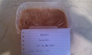
biohazard.jpg?w=300
Biohazard: mouldy soup

The volume of air in the container was measured:

Depth from clingfilm to surface of soup: 3.0 cm

Width of container: 13.5 cm

Length of container: 18.5 cm

Volume    = 3.0 cm x 13.5 cm x 18.5 cm

= 749.25 cm3

= 7.5 x 10⁻⁴ m3 (2 significant figures) Note: the corners of the container were rounded, so this figure is approximate.

The first mould began to appear on June 4 and were tiny white spots (about 1 mm in diameter), mostly around the edges of the tub.

On June 5, the white spots had grown to a diameter of around 3 mm to 4 mm, with 1 mm green/blue patches in places. More areas of growth had appeared. Condensation also appeared on the clingfilm, which made observations a little awkward.

My mould was respiring! I was so proud. My very own baby mould; they grow up so fast.

By June 9, no more spots were appearing. And it's probably a good thing, because it was getting a little crowded in there, and the patches were beginning to fight among themselves. I did NOT want to have to step in there and break anything up.

Results
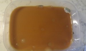
fungal-particles.jpg?w=300
The Mould Boyz

Seventeen separate areas of mould were counted. Most of the mould was clinging to the edges of the soup on the container, with a few patches in the centre. The patches in the centre resembled blisters lying just on or beneath the surface. They were milky in appearance, and slightly jelly-like, measuring 0.5 cm to 1.0 cm in diameter. Delicious.

The patches around the edge were either white, or white with green/blue areas. The white patches had stalks, while the green/blue mould was furry in texture. These patches measured around 1.5 cm to 2.5 cm across. I've seen this furry mould before; it normally inhabits the bit of sandwich you've just put into your mouth. You know this, because there's half a patch of mould left when you look at your meal.

Analysis of results

Each area of growth probably represents one fungus, which arose from one fungal spore. My result was: 17 fungal spores per 7.5 x 10⁻⁴ m3 air.

To find the number of fungal spores per cubic metre of air:

= fungal spores per m3

= 2.3 x 10⁴ fungal spores m-3 (2 significant figures)

I could work out the total number of fungal spores in the air in my whole kitchen; but frankly, I'd rather not know! I'm quite happily living in blissful ignorance, and perpetuating the dastardly rumour that I am, in fact, a great wife who cooks, cleans, maintains her rather great bottom AND makes interesting conversation that does not involve mould.

Critical thinking

The density of fungal spores I obtained is almost certainly an underestimate of the true density. This does not make me happy. I thought 22,666 spores per cubic metre was quite alarming enough.

Assumptions were made that the number of fungal spores in the air are evenly distributed throughout the room; this is unlikely to be the case, especially with movement of air. It may be that not all the spores trapped in the container grew into patches of mould. It was also assumed that all spores had grown into mould when I ended the experiment; this may not be the case.

Further investigations

We were supposed to think about what else we could investigate. But really, the only thing that came to mind was the mating habits of fungus. I suspect I've been staring at a screen for too long...

My next activity involves researching Leontopithecus rosalia. Stay tuned!

Permalink 1 comment (latest comment by David Smith, Friday, 10 June 2011, 12:47)
Share post

This blog might contain posts that are only visible to logged-in users, or where only logged-in users can comment. If you have an account on the system, please log in for full access.

Total visits to this blog: 361662